Swiss researchers create artificial lung to study Covid-19 blood clots
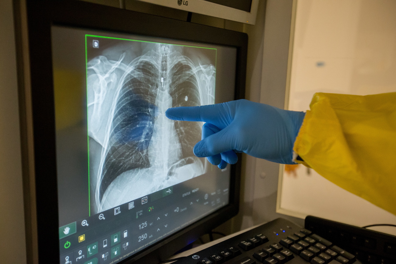
Scientists at the Federal Institute of Technology Lausanne (EPFL) have created an artificial lung to better understand how the new coronavirus causes blood clots in certain patients.
A recent studyExternal link revealed that around 10% of hospitalised Covid-19 patients develop blood clots, however, it is unclear why the virus causes this reaction. In the most severe cases, the blood clots can result in a stroke.
To investigate this phenomenon and other Covid-19 infections, EPFL scientists have developed a microfluidic chip that models the human lung and replicates part of its structure.
The chip holds lung epithelial cells, blood-vessel cells and immune-system cells, and lets scientists directly observe how the virus attacks human cells and triggers the formation of blood clots.
When that happens, scientists say two mechanisms may be occurring. One is an excess production of cytokines, which are proteins that play a role in immune-cell signalling, leading to so-called “cytokine storms”. These can damage blood vessels and cause blood clots to form, and are potentially fatal.
The other possible reason is damage to the interior lining of blood vessels – or the endothelium – in the lungs. The lungs have a lot of this kind of tissue, and when it’s damaged, blood can coagulate easily and form clots.
Lung-on-a-chip
To help find out, the EPFL team took a lung-on-a-chip device and adapted it to model the individual steps in a SARS-CoV-2 attack on the lungs. The device contains microfluidic channels that feed nutrients to cells on the chip, which are arranged to recreate a section of the lungs.
Inside the chip there is a layer of epithelial cells, the cells coating the lungs, and a layer of endothelial cells, the cells lining the blood vessels. These two layers are separated by a membrane.
During their tests, when the virus was introduced into their device it first attacked the outside layer of epithelial cells, just like in a natural infection. The team found that within a day the virus had reached the inner layer of endothelial cells and caused considerable damage over subsequent days.
“There was enough damage to destroy the endothelium and expose blood in the vessels to air, causing clots to form,” says Vivek Thacker, a postdoctoral researcher. “With our lung-on-a-chip system, we found that the virus may be causing blood clots by attacking the endothelium directly. However, that doesn’t mean that cytokines don’t play a role too and make things worse.”
To further their understanding, the team plan to use their lung-on-a-chip with actual blood samples so that they can observe clot formation directly.
More
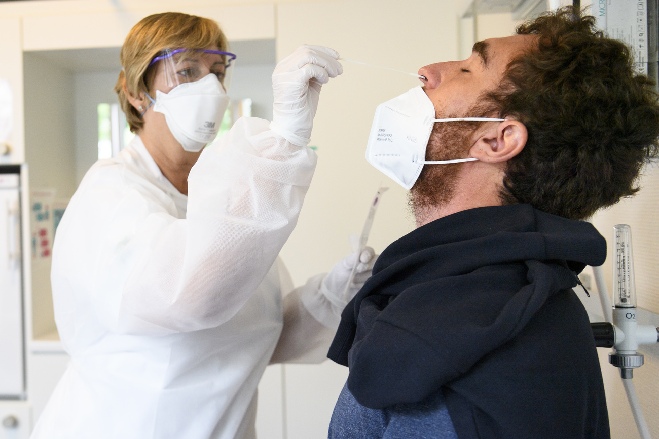
Coronavirus: the situation in Switzerland

In compliance with the JTI standards
More: SWI swissinfo.ch certified by the Journalism Trust Initiative


You can find an overview of ongoing debates with our journalists here. Please join us!
If you want to start a conversation about a topic raised in this article or want to report factual errors, email us at english@swissinfo.ch.