Award-winning photos show the hidden beauty of Swiss research
From the transparent belly of a frog to regenerating the head of a decapitated animal, the winners of the 2023 Swiss National Science Foundation’s scientific image competition show researchers’ daily routines from unusual perspectives.

When we think scientists’ everyday work, the scenes that come to mind are often people in lab coats, repeating experiments, or giving lectures heavy on scientific buzzwords. But scientists are also sharp, curious observers able to ascribe beauty and meaning to their study subjects. The best photographs taken by scientists at Swiss universities and research institutions give us a glimpse of this world.
An international jury assembled by the Swiss National Science Foundation (SNSF) awarded the winners of its annual Science Image CompetitionExternal link on April 25 in the Swiss capital Bern.
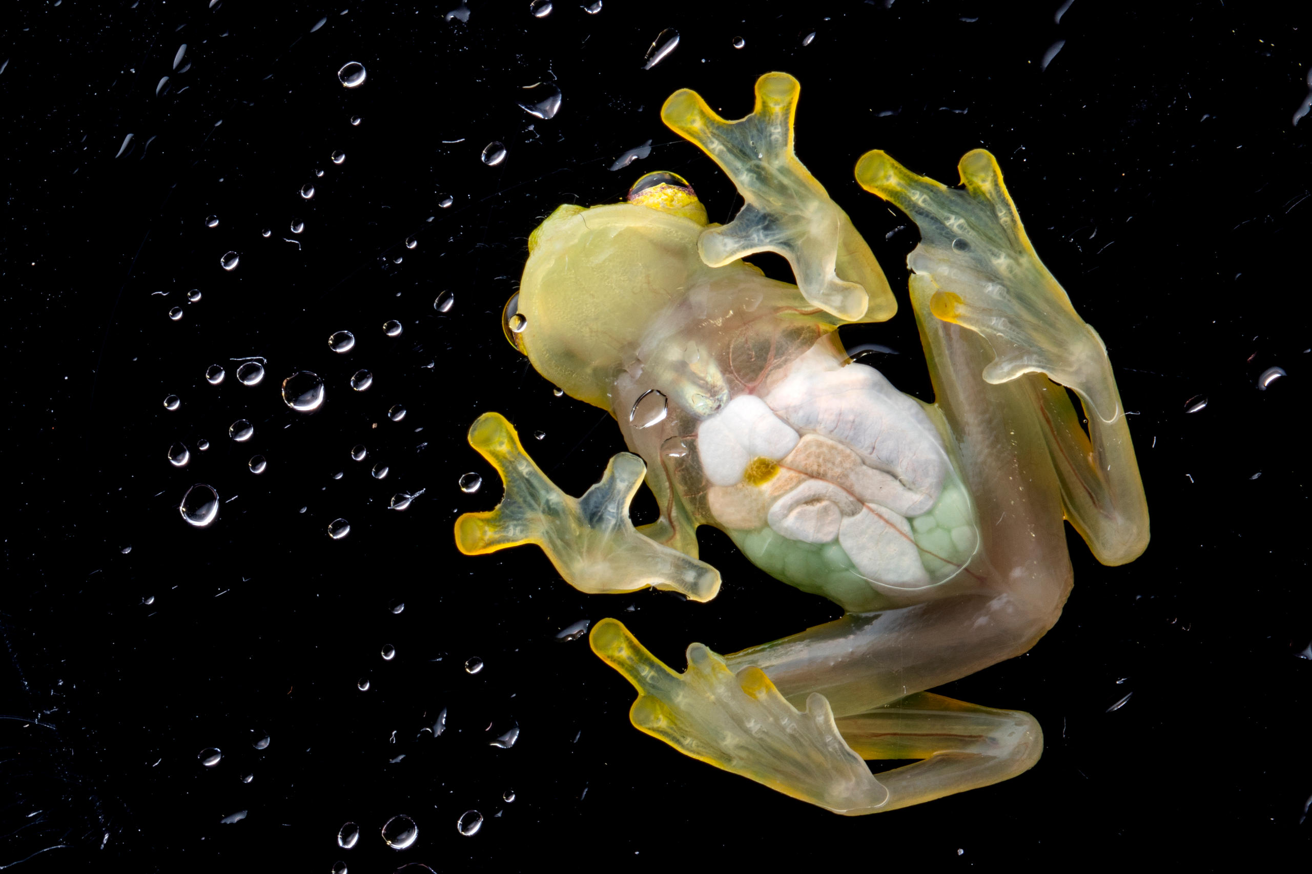
First place in the “Object of study” category was awarded to Francesca Angiolani-Larrea, a PhD student at the University of Bern who studies the ecology and evolution of amphibians, for her photo of a frog belly.
Parental care in the animal world takes diverse forms in amphibians. Case in point is Hyalinobatrachium valerioi, a type of glass frog with a transparent belly. The mother’s only tasks are to select the best mate and to produce the eggs, while fathers stay with their young and look after them – sometimes up to seven clutches of eggs at the same time.
For this photo, Angiolani-Larrea resorted to a common piece of laboratory equipment – a transparent Petri dish – to show her study subject from a “striking new perspective”, according to the jury.

Mariafrancesca Petrucci, a PhD student in veterinary medicine at the University of Bern, won first prize in the category “Women and men of science”with her self-portrait next to a miniature pig while listening to its heart. She says that the research project she conducted on pain and its characteristics in animals made her realise that “the life and health of animals, humans and other living beings are equally important, and that the highest standard of care must be guaranteed for all, wherever they are.”
Her caring gesture and the warm colours in the picture are “a soft contrast to the often-controversial topic of animal testing,” according to the jury. It “brilliantly expresses emotions that are rarely associated with science”.
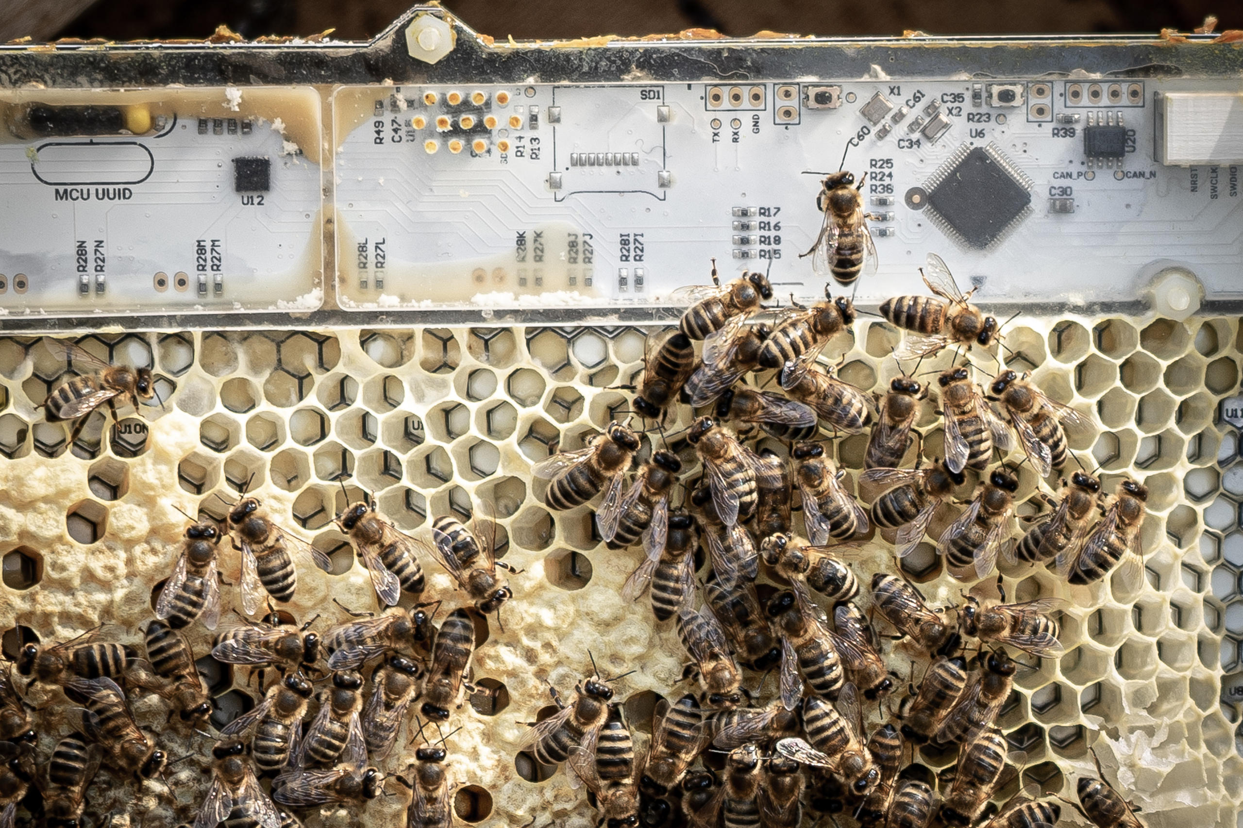
In the “Locations and instruments” category, Rafael Barmak, a doctoral assistant at the Swiss Federal Institute of Technology Lausanne (EPFL), won over judges with a close-up of a bee colony residing on a robotic device, which can interact with the bees. This helps scientists investigate their behaviour, potentially supporting them in an increasingly hostile environment.
The picture “transports us into a new biohybrid world where the natural meets the artificial,” the jury commented.
The winner of the category “Video loops” is a visualisation of the vortexes created by an aircraft wing filmed by Cyprien de Sepibus, a PhD student at the Geneva School of Landscape, Engineering and Architecture (HES-SO) and EPFL. The video impressed the judges by “magically making the invisible visible”.
The jury also awarded fifteen distinctions, more than ever before. Here are some examples:

This labyrinth-like photo of a supercomputing simulation reveals the inner world of the human vision system and its complex organic structure, showing how cerebrospinal fluid flows around the optic nerve.
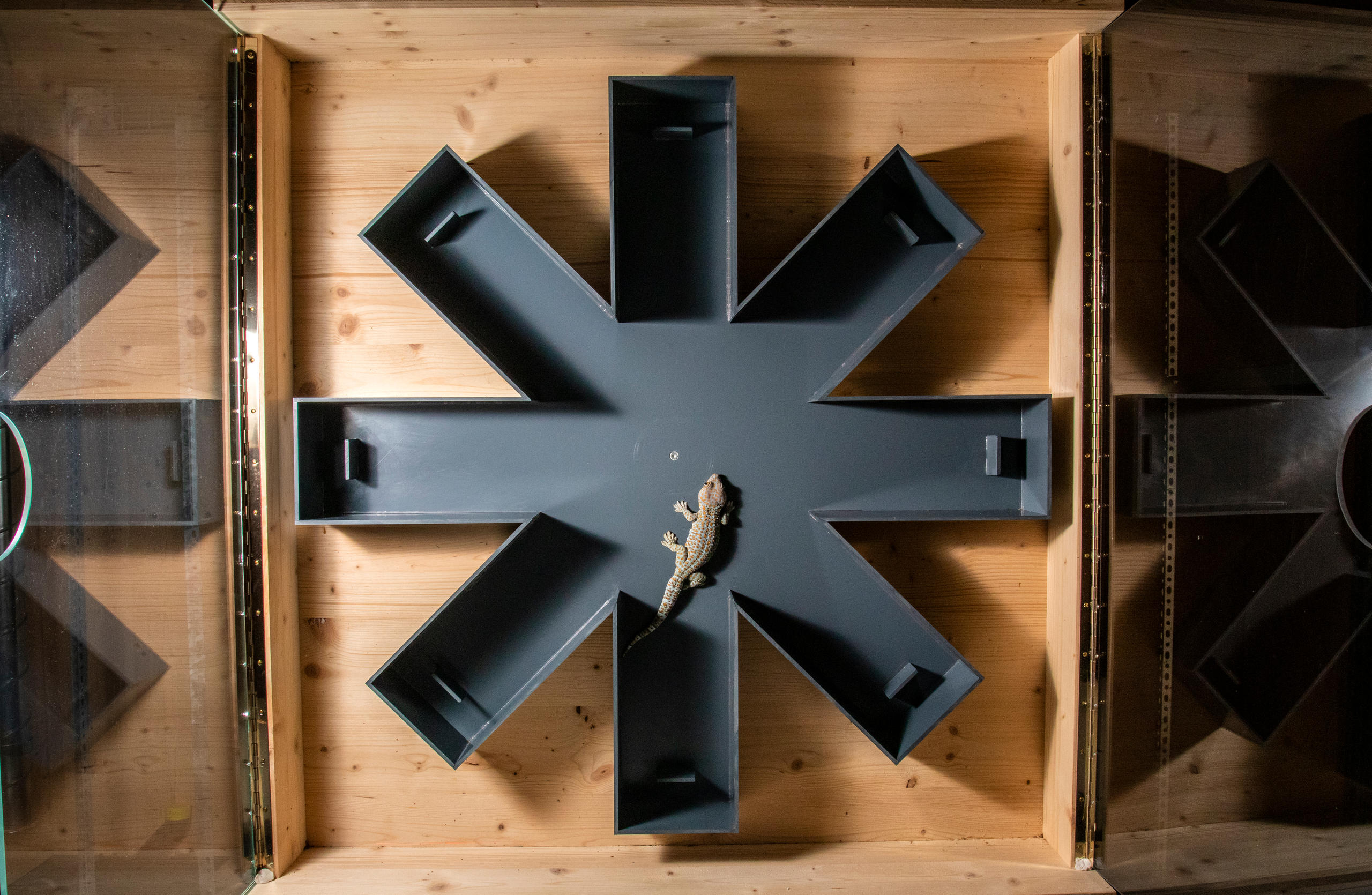
Angiolani-Larrea and her colleagues are studying how sociality and ecology influence the learning abilities of Tokay geckos. In the experiment, a gecko has to learn the location of food in a maze placed either vertically or horizontally.
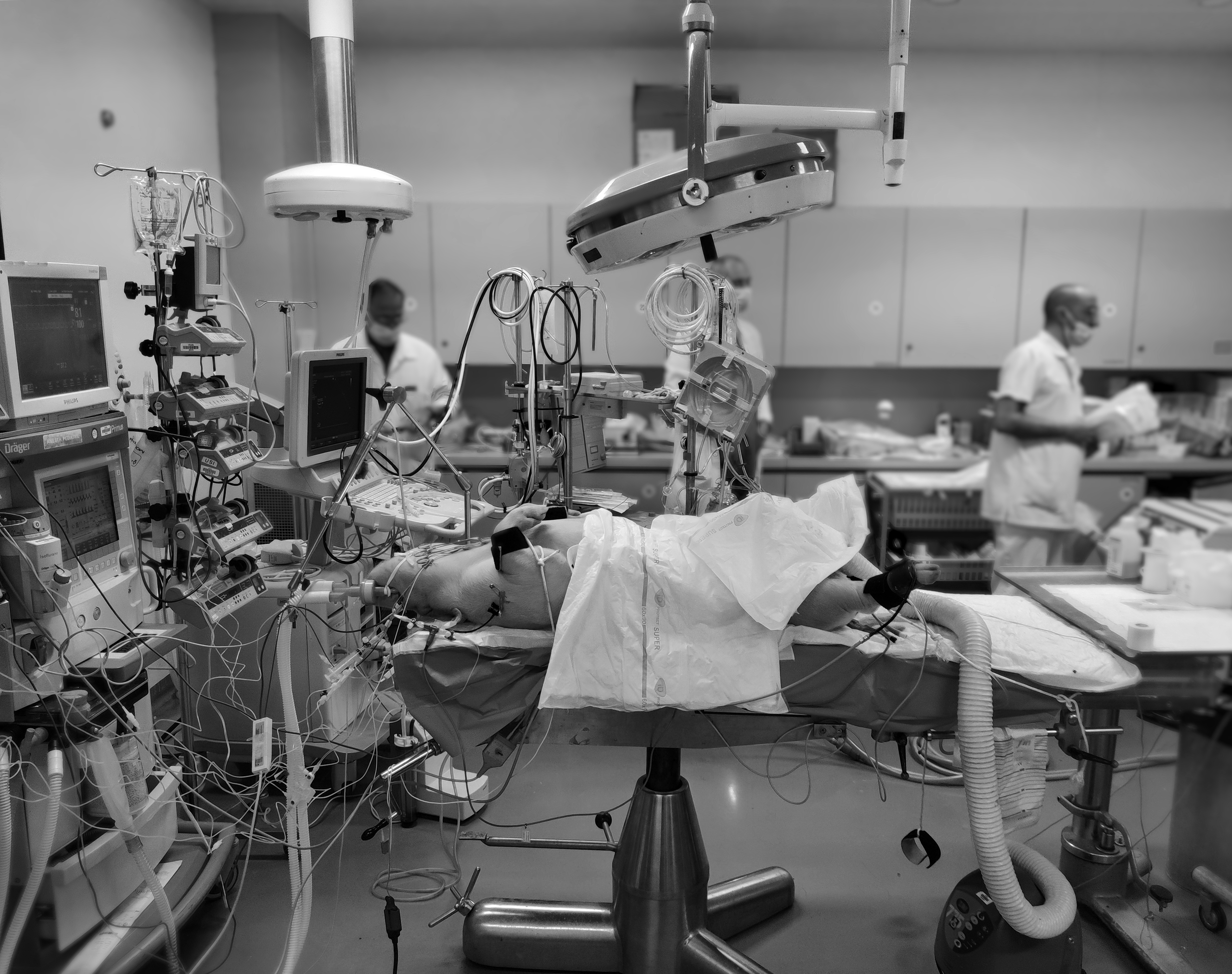
This photo was taken before the start of an operation on a pig, meant to test whether a novel blood vessel graft could improve patients’ life expectancy and quality of life. The pig lies where we would expect a human patient to be, underlining the animal’s sacrifice for improved human healthcare.
Distinctions in the “Video loops” category: “Made myself a new head” by Yamini Ravichandran, Postdoctoral researcher, University of Geneva.
This video follows the regeneration of the head of a decapitated small adult hydra over the course of four days. The resilience of this humble creature holds the promise of for advances in regenerative medicine.
Distinctions in the “Video loops” category: “Tumour growth simulation” by Roman Vetter, Senior scientist, ETH Zurich.
The animation shows a malignant tumour growing from a single cell to a million cells which reflects a striking and uncontrollable growth of cancerous proliferation.
All the images and videos submitted for the competition can be viewed in this online galleryExternal link.
The winning pictures and videos of this year’s competition can also be seen at an exhibition at the Biel/Bienne Festival of photographyExternal link running from May 5 to May 28.
Edited by Sabrina Weiss and Veronica DeVore

In compliance with the JTI standards
More: SWI swissinfo.ch certified by the Journalism Trust Initiative
You can find an overview of ongoing debates with our journalists here . Please join us!
If you want to start a conversation about a topic raised in this article or want to report factual errors, email us at english@swissinfo.ch.