Can neuroscientists read our minds?
Powerful imaging technology has led to huge advances in understanding how our brains work. But should we be worried about opening our minds to outside scrutiny?

Neuroscience experts took part in a roundtable talk in Geneva on Wednesday as part of Brain Week to discuss the progress made in unlocking the secrets inside our heads and the ethical implications.
Brain Week has taken place for the past 12 years in Europe, organised by the European Dana Alliance for the Brain. The association’s goal is to help the public understand the importance of this type of research. Events are being organised in seven cities across Switzerland between March 16-21.
Over the past 15 years neuroscience has witnessed spectacular developments, benefitting from much more powerful and precise magnetic resonance imaging (MRI), Patrik Vuilleumier, director of Geneva University’s Neuroscience Centre, told the 300-strong audience at Geneva University.
“Huge progress has been made in understanding the specific functions of different brain regions, but I don’t think we have a great deal to fear at the moment,” said Vuilleumier.
Although neuroscientists can now measure brain activities linked to different kinds of memories, technology is still a very long way off decoding and extracting our innermost thoughts, he explained.
Exploratory work
Most regions of the brain have now been explored and are well documented.
We now have a better understanding of how the human visual system recognises objects, patterns and faces, how we identify sounds and voices, and how we perceive and respond to emotions and social signals, such as facial expressions or eye gaze.
One area of research enjoying huge growth is the classification of MRI pixel patterns, based on the kind of images shown.
In March 2008 researchers from California University created a model for decoding brain information. They managed to identify a specific image that a patient was looking at among dozens of others simply by observing their brain activity.
Several months later Japanese neuroscientists went even further by first identifying an image observed by someone lying in an MRI scanner, and then rebuilding the image on a screen – but in simple geometric shapes.
Sound and space
Research into decoding brain responses to sounds and voices is also becoming much more sophisticated, said Vuilleumier.
“It’s possible to differentiate between different people talking, the vowel sounds they use and their intonation, whether they express anger or sadness,” he said.
And earlier this year British scientists at University College London published a study into spatial memory, where they were able to accurately predict someone’s position within a virtual environment solely from the pattern of activity in a part of the brain called the hippocampus.
The study was part of an investigation aimed at learning how memories are created, stored and recalled. The research prompted the British team to call for an ethical debate on how brain imaging may be used in the future, and the kind of safeguards that need to be put in place to protect people’s privacy.
Ethical questions
Ethical discussions in the neuroscience community are ongoing, particularly over responsibility and free will, said Vuilleumier.
“The biggest ethical question at the moment is misuse,” said Vuilleumier. “The risk is that people over-interpret what we can measure.”
There have been recent cases in the United States where lawyers have asked in court for a client to have a brain scan to assess their emotional responses.
But the science is not at that stage yet, he said. The limitations of imaging technology are essentially down to the complexity and speed of our brains.
“With most MRI techniques you measure changes in energy in different 2mm pixels, or points in the brain, where there are almost a million neurons and each neuron can contribute to an aspect of our mental state. So it’s not possible to decode very precisely,” he said.
And for now, researchers can only measure relatively slow changes in brain activity – 1-5 seconds long.
“Thoughts are much faster,” said Vuilleumier.
Not mindreading
Alexandre Mauron, professor of bioethics at Geneva University, also tried to reassure the audience, saying recent developments had almost nothing to do with mindreading.
“Mindreading is a fantasy; the idea that one day a machine could burst into our universe – our internal cinema – and discover our innermost thoughts. This fantasy is terrifying as we have a tradition of freedom of thought that so far no one has been able to touch up to now, even in the worst totalitarian regimes,” he said.
The problem is that our culture has been marked by a Cartesian dualism that separates our body – and brain – from our soul, or thoughts. It is very difficult to detach ourselves from this way of thinking, he said.
“Our ‘folk psychology’, our normal way of talking about our thoughts, emotions, mental state and choices, is a language that is increasingly out of touch with the language of neuroscience,” he said.
swissinfo, Simon Bradley in Geneva
Switzerland has a fast-growing neuroscience community: the Swiss Society for Neuroscience has more than 1,000 members.
There is close cooperation and interaction between Geneva and Lausanne universities and university hospitals, and the Federal Institute of Technology, Lausanne, with more than 80 neuroscience research groups.
Zurich also has a joint neuroscience centre creating synergies between some 440 neuroscientists, or 100 research groups, at the Federal Institute of Technology, Zurich, and Zurich University.
Switzerland was chosen to host the sixth Forum of European Neuroscience based on the strength of its neuroscience research and infrastructure.
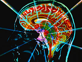
Nuclear magnetic resonance imaging (MRI) is primarily a medical imaging technique most commonly used to visualise the structure and function of the body.
MRI is especially useful when it comes to differentiating between the different soft tissues of the body, and is often employed for neurological, musculoskeletal, cardiovascular and cancer imaging.
The technique involves a powerful magnetic field that aligns and rotates hydrogen atoms found in water in the body. The signal they emit can be manipulated so as to reconstruct an image of the body.
Diffusion MRI uses the properties of water molecules in the body. For example, they are more likely to move along the axis of a neural fibre, giving its direction.
Functional MRI measures blood flow to determine when an area of the brain is activated for example, but does not show the underlying network.


In compliance with the JTI standards
More: SWI swissinfo.ch certified by the Journalism Trust Initiative


You can find an overview of ongoing debates with our journalists here . Please join us!
If you want to start a conversation about a topic raised in this article or want to report factual errors, email us at english@swissinfo.ch.